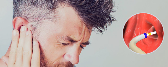


The device is guided through the nasal passageway until it reaches the eustachian tube opening in the back of the nose. Under direct visualization, the balloon is then inserted into the Eustachian tube and inflated to dilate the blocked tube — similar to the way a stent is used to open up a clogged artery.
Before the procedure, your surgeon will place a small endoscope — a thin tube with a camera and light — through the nasal opening to examine the nasal cavity and look for any potential causes of ETD or assess for anatomical issues that may impact the treatment setting for you. Examples of these include ruling out sinus infections, extent of acid reflux, severity of allergic rhinitis, presence of septal deviations, etc.
On the day of your procedure, an endoscope is introduced into the nose with the deflated Eustachian tube balloon. The device is guided through the nasal passageway until it reaches the eustachian tube opening in the back of the nose. Under direct visualization, the balloon is then inserted into the Eustachian tube and inflated to dilate the blocked tube — similar to the way a stent is used to open up a clogged artery. The balloon is kept inflated for about two minutes helping to remodel the Eustachian tube cartilage and flatten out any inflammation. The balloon is then deflated and removed from the Eustachian tube. In the case that both Eustachian tubes are affected, the procedure can be performed on the opposite side as well.
For decades, the gold standard of treatment for patients with chronic eustachian tube dysfunction has been repeated placement of ear tubes. However, these tiny plastic cylinders that are surgically placed in the ear can fall out over time and long-term use can lead to perforation and often necessitates dry ear precautions. Since Eustachian Tube Balloon Dilation was introduced in 2010, numerous studies have shown the procedure has several distinct benefits.
Improved outcomes: In addition to draining fluid and equalizing the pressure in the eustachian tube, using a balloon to dilate the passageway can also help address the underlying structural problem. Studies show the procedure causes cellular changes that allow the connective tissues to remodel
Reduced need for repeat surgeries: Research has found patients experience long-lasting relief with Eustachian Tube Balloon Dilation when compared to tubes, which often may need to be replaced several times due to them falling out and can lead to scarring or holes developing that subsequently need to be repaired
Safety: The procedure can be performed in a doctor’s office with local anesthesia and numbing agents, rather than in the operating room under general anesthesia. If the patient does not think they can tolerate the procedure or has other complications, a traditional surgical setting can be arranged
Minimal recovery time and follow-up care: Balloon dilation is well tolerated. Patients are typically back to work and take part in normal activities often within the same day of the procedure and experience very mild side effects. Nasal packing is not usually needed.